Preventing Rectus Diastasis

7 Tips for a Strong Core
Ever heard of a mummy tummy or mom pooch?
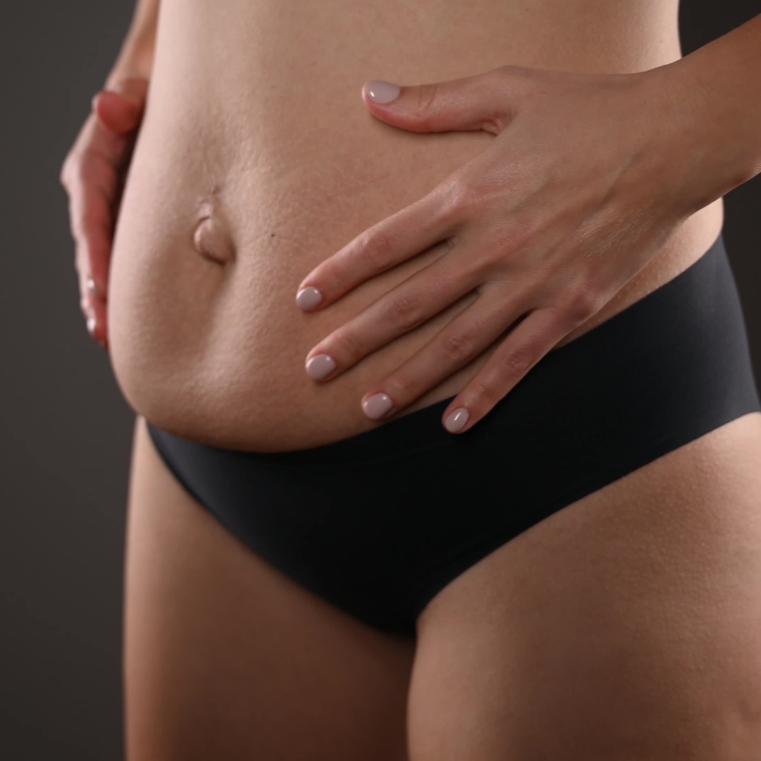
Some women continue to look pregnant months or years after giving birth. This term is clinically called a number of things: abdominal separation, rectus abdomins diastasis, rectus diastasis, diastasis recti. All these terms are abbreviated as DRA. The traditional determinant for a DRA is a 2 finger width separation recorded above, at, and below the umbilicus.
Why does it happen?
The abdominal muscles separate to accommodate for the growing baby and increased pressure on the midline. This condition affects almost every single mother after childbirth. For many, it resolves with little intervention. So how do you know if you need to do something more about it?
Why is screening important?
The pelvic muscles and the abdominal muscles work together to maintain abdomino-pelvic control in functional tasks. DRA results in impaired abdominal muscle and pelvic muscle strength and stability.
A DRA may affect posture, trunk stability and motion, respiration, urination, and vaginal delivery. Strengthening of the transversus abdominis muscle in isolation and with functional activities is key to resolving a DRA and improving core stabilization.
How do I mitigate risk?
1. Engage in Safe Core Exercises
Focus on exercises that engage the deeper abdominal muscles, such as pelvic tilts, modified planks, and bridges, while avoiding traditional crunches and sit-ups that often cause increased strain along the midline.
2. Good Posture
How your body is stacked dictates where the stress points lie within the body. Stand and sit tall, engaging your core muscles to support your spine and abdomen, reducing unnecessary pressure on the midline.
- Excess forward shoulders and hunching disengages the core muscles and places more stress on the back.
- Excess backward bending of the spine will increase stress through the lower back in particular leading to risk of overuse injury.
3. Lifting Techniques
This idea circles back to maintaining good posture. Improper lifting techniques will place excess stress on areas like the spine leading to an increased risk of injury.
- Lift with your legs with a wide base of support.
- Keep the weight close to your body.
- Engage the core muscles to support the back/spine.
Improper lifting can exacerbate diastasis recti and this is shown through doming of the abdominals.
4. Supportive Belly Binders or SI Belts
These should be a consideration for anyone during the later stages of pregnancy, especially those that report back and pelvic pain. Speak to your physical therapist for an assessment to see which one would be more effective for you. Most clinics have samples that you can try on and test to see if your pain reduces and stability improves while wearing one.
5. Gradual Weight Gain During Pregnancy
Maintaining a healthy and gradual weight gain during pregnancy can help minimize the pressure on the abdominal muscles, reducing the risk of separation.
6. Be Mindful of Your Breathing
Proper breathing techniques are crucial for preventing diastasis recti. Focus on diaphragmatic breathing, which involves expanding the belly as you inhale and gently drawing the belly button towards the spine as you exhale, supporting the core muscles.
7. Ask for Help
Consulting with a certified fitness trainer or physical therapist with expertise in diastasis recti prevention can be highly beneficial. They can create a personalized exercise plan and check to make sure you are engaging your inner core muscles during exercises. This can be done manually or with real-time ultrasound.
Treatment Options for DRA
Manual therapy techniques used to treat patients with DRA include myofascial release, trigger point release, muscle energy techniques, and visceral manipulation. Other manual therapy techniques used include manual facilitation for abdominal contraction, soft tissue mobilization, connective tissue manipulation, and joint mobilization at the sacrum, innominate, lumbar spine, coccyx, and pubic symphysis. Rehabilitative Ultrasound Imaging may also be used to assist in treatment to assist with visualization and activation of the correct muscles.


